- Empty cart.
- Continue Shopping

Recombinant Anti-PD-L1 antibody [28-8]
Recombinant Anti-PD-L1 antibody [28-8]
This product is currently out of stock and unavailable.
Key features and details
- Produced recombinantly (animal-free) for high batch-to-batch consistency and long term security of supply
- Rabbit monoclonal [28-8] to PD-L1
- Suitable for: ICC/IF, IHC-P, WB, Flow Cyt, IHC-Fr
- Knockout validated
- Reacts with: Human, Recombinant fragment
Overview
Product name
See all PD-L1 primary antibodies
Description
Host species
Tested applications
Species reactivity
Immunogen
Recombinant full length protein corresponding to Human PD-L1 (extracellular). The immunogen contains the specific extracellular domain of huPD-L1 (Phe19-Thr239). See reference for more info – www.ncbi.nlm.nih.gov/pmc/articles/PMC4561627/
Database link: Q9NZQ7
Positive control
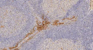
- Tissue: Human tonsil, head and neck squamous cell carcinoma and placenta tissues; L2987 cell line. Cell Lines: Positives: B-CPAP (high), ES-2 (medium), HCC70 (low), CHO-PDL1, U-87 MG For additional information – please refer to this publication: Programmed death-ligand 1 (PD-L1) expression in various tumor types – http://www.immunotherapyofcancer.org/content/1/S1/P53 IHC-Fr: Frozen human tonsil tissue sections
General notes
FURTHER INFORMATION ON POSITIVE CONTROLS (Chinese version)
Tissue:
– Tonsil – with hyperreactive changes. Screening of hyper-reactive tonsils is recommended to find tonsil with the highest expression of PD-L1 in crypt epithelium, macrophages homing the germinal centers and interfollicular mononuclear leukocytes.
– Tumor tissues – prescreened for positive tumor and inflammatory infiltrates. PD-L1 expression varies by tumor type so screening is recommended to find positive and negative tumor controls.
– The following publication is useful for finding suitable tumor types for PD-L1 expression:
http://www.immunotherapyofcancer.org/content/1/S1/P53
– Note: Look for specimens with high numbers of inflammatory macrophages and mononuclear leukocytes.
Cell Lines:
– Positive controls: B-CPAP (high), ES-2 (medium), HCC70 (low).
Primary antibody negative control: Recombinant Rabbit IgG isotype control antibody, ab172730.
Recombinant protein positive control: Recombinant human PD-L1 protein, ab167713.
Immunohistochemistry usage:
For IHC usage on FFPE tissues, we recommend using PD-L1 IHC panel ab236676, which contains PD-L1 antibody clone 28-8 (ab205921), HIER antigen retrieval reagent (ab208572) and IHC detection kit HRP/DAB (ab209101).
Western blot usage:
For clone 28-8, it is recommended to use Odyssey system. This system has the advantages of a wider dynamic range and less background than chemiluminescence.
This PD-L1 antibody [28-8] has been used as detector antibody in Human PD-L1 SimpleStep ELISA® kit: ab214565 and ab277712.
This product is a recombinant monoclonal antibody, which offers several advantages including:
- – High batch-to-batch consistency and reproducibility
- – Improved sensitivity and specificity
- – Long-term security of supply
- – Animal-free production
Our RabMAb® technology is a patented hybridoma-based technology for making rabbit monoclonal antibodies. For details on our patents, please refer to RabMAb® patents.
Properties
Form
Storage instructions
Storage buffer
Preservative: 0.01% Sodium azide
Constituents: 59% PBS, 40% Glycerol, 0.05% BSA
- Batch dependent within range: 50 µl at 0.994 – 1.095 mg/ml
Purity
Clonality
Clone number
Isotype
Research areas
- Immunology
- Cell Type Markers
- CD
- Non-lineage
- Immunology
- Immune System Diseases
- Autoimmune
- Cancer
- Tumor immunology
- Tumor-associated antigens
- Cancer
- Tumor biomarkers
- Tumor antigens
- Signal Transduction
- Antibodies
- pd-l1


